


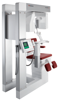
SCANORA® 3D
More value for your money
Cone Beam 3D and digital panoramic imaging combined in one system
SOREDEX has been designing and manufacturing high quality dental imaging systems for over thirty years. With SOREDEX your investment is protected, because our products are renowned for their excellent quality, total reliability and extremely long service life.
The SCANORA® 3D system makes advanced dental imaging fast and easy. We let you concentrate on your most important activity – treating your patient.
Watch SCANORA® 3D video
SCANORA® 3D
|
 |
Excellent diagnostic performance
The SCANORA® 3D system off ers a modern way of seeing dentomaxillofacial anatomy and solving diagnostic tasks. The highdefinition panoramic image shows the regions that need further investigation. The optimum 3D technique for a specifi c task can be easily selected, treatment planned and fi nally follow-up studies done, all with one effi cient unit. The system contains user-selectable features that contribute to excellent diagnostic performance.
Uncompromised quality
The SCANORA® 3D system has been designed from the ground up using the very latest 3D imaging technology. The overall quality comes as a result of successful combination of the x-ray unit itself, ergonomic and rigid patient support devices, and the newest image handling procedures to enhance diagnostic information.
High technology flat-panel detector
The flat-panel detector is a masterpiece of modern CMOS technology. Compared to older generation of image intensifiers, flatpanel detectors offer superior image quality due to their large dynamic range, better contrast and lack of image distortion. Additionally they are insensitive to electromagnetic interference, are compact in size and have a very long service life.
Full diagnostic information
The FOV can be easily positioned anywhere in the maxillofacial area, thanks to the motorized patient seat. After scanning and image reconstruction, a full range of diagnostic options can be utilized. The diagnostic information can be thoroughly examined with the many powerful software tools and features.

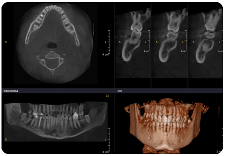
The small FOV is for localized problems.
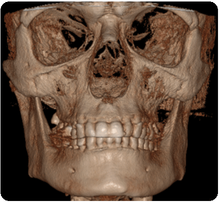
The optional XL FOV is suitable, for instance, in sinus examinations.
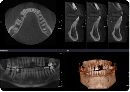
The medium FOV can show all the teeth in one image.
An example of the large FOV.